|
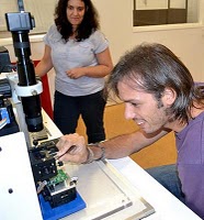
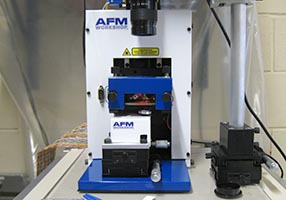
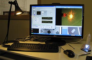
New Instrument In early january 2019, we installed our second TT-AFM instrument from AFMWorkshop. We now have two fully independent AFM instruments.
|
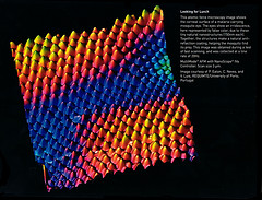
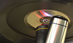
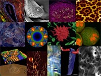
Prize Winning Image and Magazine
Cover An image I produced from some work in collaboration with Ricardo Franco of
UNL, and Thomas Hanscheid and Carolina Tempera of IMM, won the
2nd Prize in the category Electron Microscopy - Physical
Science, in the Royal Microscopical Society 2014 Scientific Imaging
Competition. This image was recently featured on the cover of the RMS
Magazine (shown right). The image was titled Field of Petals, and shows an SEM
image of hemozoin-like crystals secondary electron image measured at 15
kV. Image represents a magnification of ca. 5000x, the horizontal field
width is 55 microns. These crystals are part of a project seeking to
develop new antimalarial compounds. Sample provided by Carolina Isabel
Glória Tempera, of the Instituto de Medicina Molecular, Lisbon.
|

|
Our Work on Tropical Diseases in the News
A news story about our work against leishmaniasis using frog
peptides has been featured here
on the AFMWorkshop site.
The story is related to our collaboration with the group from UFPI in
Brazil. The correct link to access the paper discussed is: http://dx.doi.org/10.1016/j.nano.2013.09.003.
In related news, we are glad to welcome José Roberto Leite from UFPI to
the group as a post-doc during 2015.
|
|
Special Visiting
Researcher
I was made a "Pesquisador Visitante Especial" - Special Visiting
Researcher at the Federal University of Parnaíba, Brazil. This will
probably run from 2015 to 2018. The grant is funded by CNPq (Brazil).
Among other things (I'll be in Brazil a lot!), we will have funding
soon for a post-doc in Brazil,
and a grant for a PhD sandwich student between Brazil and Portugal.
The themes will be natural products, nanoparticles and AFM. Contact me
if you are interested in either of these positions. See this
news piece on the UCIBIO website for more details.
|
|
Well done
Cristina!
Cristina Neves has successfully defended her PhD. Thesis, which was
titled "Development of fluorescent silica nanoparticles encapsulating
organic and inorganic fluorophores: synthesis and characterization"?.
Congratulations to Cristina, who was awarded the degree with
distinction due to her hard work and excellent defence.
|
|
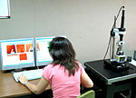
New AFM Installed
We have just installed a new instrument in the lab. It is an
LS-AFM
from AFMWorkshop. This is the first instrument of its kind in the
world, and is a new atomic force microscope designed for life sciences
applications. The instrument allows optical microscopy (including phase
contrast microscopy), epifluorescence and AFM on the same sample. It
also allows observation of the sample from above (reflected light), or
below (transmitted light). It is ideal for examination of cells.
Initial tests last week suggest the new instrument is going to have
excellent performance. Optimisation and a more complete installation is
ongoing.
This instrument forms part of the BIO-AFM network which means that
any researchers who wish to use it should be able to. Please contact me
via the email address at the bottom of the page if you are interested. |
 |
Fellow of the
Royal Microscopical Society
I was elected to be a Fellow of the Royal Microscopical Society (FRMS).
Thanks very much to the RMS for
this honour. |
|
Cristina
wins Nano2012 and E-MRS Young Scientist 2013 Prizes
Congratulations to Cristina Neves, my PhD student, on
winning a Young Scientist Award at the recent
E-MRS
Spring
Meeting. She won the award for her presentation titled
"Fluorescent Silica Nanoparticles based on Organic and Inorganic
Fluorophores: Preparation and Characterization". This is
the second
prize Cristina has won for her work in the
project on fluorescent nanoparticles. Cristina will soon be writing up,
so if any other prospective PhD student are interesting in continuing
work on this award-winning, FCT-funded project, they should contact me
ASAP! Once again, congratulations for Cristina, and we hope to see this
great work written up in thesis form really soon! |
|
Requimte
AFM Workshop 2013
The AFM Workshop
here in Porto took place last week. The course was a great success and
we had lots of positive feedback. There's a blog post reviewing the
course here:
http://atomicforceblog.blogspot.pt/2013/04/requimte-afm-workshop-2013.html
|
 |
Requimte AFM
Workshop Prize
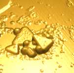
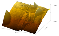
Congratulations
to Leonor Soares who won the image processing
competition in the Requimte AFM workshop 2013, with her image "Leonor
hair details", which is shown here. Leonor prepared (and grew!) the
sample herself, and scanned it along with Jorge, Inés Rocha and Tiago
Galvão. She then produced this nice image which shows surface details
very well. |
 |
Announcment of
the Requimte 2013 AFM Workshop
Since the acquisition of two AFM instruments in Requimte two years ago,
we've been training various users in the technique. A successful course
was held in 2011, and now we are announcing the 2013 course.
The course will be held in our laboratory in Porto, with two
instruments for the students to use. The course will include 2 days of
theory and two days of practice, covering both Image acquisition and
data analysis. The course will take place between the 25th and the 28th
of March 2013. Places are limited, so interested students are
encouraged to reserve a place as soon as possible.
Visit afmhelp.com/requimte
for more information. Reservations can be made by emailing
afmhelp@gmail.com
|
 |
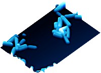
Malaria
Paper
We have published a new paper relating some studies we've made of the
development of the malaria parasite, plasmodium within liver cells
(hepatocytes), using AFM. This is the first time anyone has ever
studied the parasite in liver cells using AFM. In fact, there have only
been a couple of studies of hepatocyte sat all. Our work reports not
just AFM imaging but also nanomechanical measurements of the infected
cells.
The results show that it's possible to observe the growth and
multiplication of plasmodium within the parasitophorous vacuole (PV) as
it grows, and that this affects cell membrane texture.
But more importantly, we saw dramatic and significant changes in cells
stiffness in infected cells. 48 hours after infection, the cells had
stiffened by approximately ten times. This effect was seen in the main
body of the cell, away from the location of the PV. This suggests that
it is a reaction of the cell to the presence of the parasite. this is
the first time that cell stiffness changes upon malaria infection have
been seen in hepatic cells. The paper has been published in
Nanomedicine: Nanotechnology, Biology and Medicine, and can be found
at:
http://www.sciencedirect.com/science/article/pii/S1549963411003777
There is also a local copy for download here: eaton_etal_malaria_nanomedicineNBM_reviewed.pdf
This work was carried out at The Instituto de Medicina Molecular,
Lisbon. Many thanks to my colleagues from there, especially Nuno Santos
for allowing use of the JPK Nanowizard II AFM. The full reference is:
P. Eaton, V. Zuzarte-Luis, M. M. Mota, N. C.
Santos, M. PrudFncio, Infection by Plasmodium
changes shape and stiffness of hepatic cells Nanomedicine:
Nanotechnology, Biology and Medicine 8(1), 17-19 (2012).
|
 |


 Neves
et al, "New insights into the use of magnetic force microscopy to
discriminate between magnetic and nonmagnetic nanoparticles"
Neves
et al, "New insights into the use of magnetic force microscopy to
discriminate between magnetic and nonmagnetic nanoparticles"